scale-up from shake flasks to bioreactor, based on power input and
Anuncio
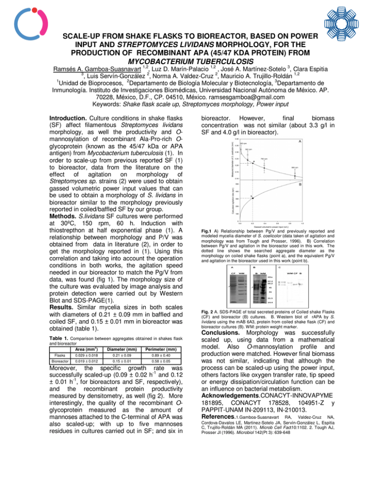
SCALE-UP FROM SHAKE FLASKS TO BIOREACTOR, BASED ON POWER INPUT AND STREPTOMYCES LIVIDANS MORPHOLOGY, FOR THE PRODUCTION OF RECOMBINANT APA (45/47 KDA PROTEIN) FROM MYCOBACTERIUM TUBERCULOSIS 1,2 1,2 3 Ramsés A. Gamboa-Suasnavart , Luz D. Marín-Palacio , José A. Martínez-Sotelo , Clara Espitia 3 2 2 , Luis Servín-González , Norma A. Valdez-Cruz , Mauricio A. Trujillo-Roldán 1,2 1 Unidad de Bioprocesos, 2Departamento de Biología Molecular y Biotecnología, 3Departamento de Inmunología. Instituto de Investigaciones Biomédicas, Universidad Nacional Autónoma de México. AP. 70228, México, D.F., CP. 04510, México. [email protected] Keywords: Shake flask scale up, Streptomyces morphology, Power input Introduction. Culture conditions in shake flasks (SF) affect filamentous Streptomyces lividans morphology, as well the productivity and Omannosylation of recombinant Ala-Pro-rich Oglycoprotein (known as the 45/47 kDa or APA antigen) from Mycobacterium tuberculosis (1). In order to scale-up from previous reported SF (1) to bioreactor, data from the literature on the effect of agitation on morphology of Streptomyces sp. strains (2) were used to obtain gassed volumetric power input values that can be used to obtain a morphology of S. lividans in bioreactor similar to the morphology previously reported in coiled/baffled SF by our group. Methods. S.lividans SF cultures were performed at 30ºC, 150 rpm, 60 h. Induction with thiostrepthon at half exponential phase (1). A relationship between morphology and P/V was obtained from data in literature (2), in order to get the morphology reported in (1). Using this correlation and taking into account the operation conditions in both works, the agitation speed needed in our bioreactor to match the Pg/V from data, was found (fig 1). The morphology size of the culture was evaluated by image analysis and protein detection were carried out by Western Blot and SDS-PAGE(1). Results. Similar mycelia sizes in both scales with diameters of 0.21 ± 0.09 mm in baffled and coiled SF, and 0.15 ± 0.01 mm in bioreactor was obtained (table 1). Table 1. Comparison between aggregates obtained in shakes flask and bioreactor 2 Area (mm ) Diameter (mm) Perimeter (mm) Flasks 0.029 ± 0.018 0.21 ± 0.09 0.89 ± 0.40 Bioreactor 0.019 ± 0.012 0.15 ± 0.01 0.58 ± 0.05 Moreover, the specific growth rate was successfully scaled-up (0.09 ± 0.02 h-1 and 0.12 -1 ± 0.01 h , for bioreactors and SF, respectively), and the recombinant protein productivity measured by densitometry, as well (fig 2). More interestingly, the quality of the recombinant Oglycoprotein measured as the amount of mannoses attached to the C-terminal of APA was also scaled-up; with up to five mannoses residues in cultures carried out in SF; and six in bioreactor. However, final biomass concentration was not similar (about 3.3 g/l in SF and 4.0 g/l in bioreactor). Fig.1 A) Relationship between Pg/V and previously reported and modeled mycelia diameter of S. coelicolor (data taken of agitation and morphology was from Tough and Prosser, 1996). B) Correlation between Pg/V and agitation in the bioreactor used in this work. The dotted line shows the searched aggregate diameter as the morphology on coiled shake flasks (point a), and the equivalent Pg/V and agitation in the bioreactor used in this work (point b). Fig. 2 A. SDS-PAGE of total secreted proteins of Coiled shake Flasks (CF) and bioreactor (B) cultures. B. Western blot of rAPA by S. lividans using the mAB 6A3, protein from coiled shake flask (CF) and bioreactor cultures (B). WM: protein weight marker. Conclusions. Morphology was successfully scaled up, using data from a mathematical model. Also O-mannosylation profile and production were matched. However final biomass was not similar, indicating that although the process can be scaled-up using the power input, others factors like oxygen transfer rate, tip speed or energy dissipation/circulation function can be an influence on bacterial metabolism. Acknowledgements.CONACYT-INNOVAPYME 181895, CONACYT 178528, 104951-Z y PAPPIT-UNAM IN-209113, IN-210013. References.1.Gamboa-Suasnavart RA, Valdez-Cruz NA, Cordova-Davalos LE, Martinez-Sotelo JA, Servín-González L, Espitia C, Trujillo-Roldán MA (2011). Microb Cell Fact10:1102. 2. Tough AJ, Prosser JI (1996). Microbiol 142(Pt 3): 639-648
