transmural, can be treated with intubation, with the tube inflated
Anuncio
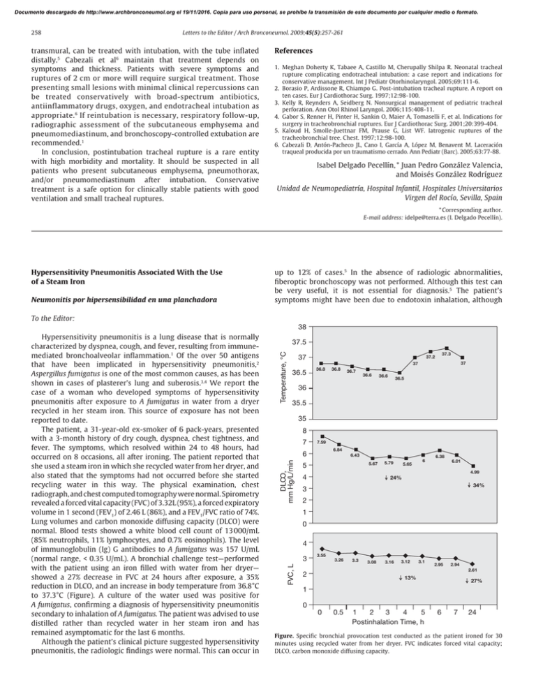
Documento descargado de http://www.archbronconeumol.org el 19/11/2016. Copia para uso personal, se prohíbe la transmisión de este documento por cualquier medio o formato. 258 Letters to the Editor / Arch Bronconeumol. 2009;45(5):257-261 transmural, can be treated with intubation, with the tube inflated distally.5 Cabezali et al6 maintain that treatment depends on symptoms and thickness. Patients with severe symptoms and ruptures of 2 cm or more will require surgical treatment. Those presenting small lesions with minimal clinical repercussions can be treated conservatively with broad-spectrum antibiotics, antiinflammatory drugs, oxygen, and endotracheal intubation as appropriate.6 If reintubation is necessary, respiratory follow-up, radiographic assessment of the subcutaneous emphysema and pneumomediastinum, and bronchoscopy-controlled extubation are recommended.1 In conclusion, postintubation tracheal rupture is a rare entity with high morbidity and mortality. It should be suspected in all patients who present subcutaneous emphysema, pneumothorax, and/or pneumomediastinum after intubation. Conservative treatment is a safe option for clinically stable patients with good ventilation and small tracheal ruptures. References 1. Meghan Doherty K, Tabaee A, Castillo M, Cherupally Shilpa R. Neonatal tracheal rupture complicating endotracheal intubation: a case report and indications for conservative management. Int J Pediatr Otorhinolaryngol. 2005;69:111-6. 2. Borasio P, Ardissone R, Chiampo G. Post-intubation tracheal rupture. A report on ten cases. Eur J Cardiothorac Surg. 1997;12:98-100. 3. Kelly R, Reynders A, Seidberg N. Nonsurgical management of pediatric tracheal perforation. Ann Otol Rhinol Laryngol. 2006;115:408-11. 4. Gabor S, Renner H, Pinter H, Sankin O, Maier A, Tomaselli F, et al. Indications for surgery in tracheobronchial ruptures. Eur J Cardiothorac Surg. 2001;20:399-404. 5. Kaloud H, Smolle-Juettnar FM, Prause G, List WF. Iatrogenic ruptures of the tracheobronchial tree. Chest. 1997;12:98-100. 6. Cabezali D, Antón-Pacheco JL, Cano I, García A, López M, Benavent M. Laceración traqueal producida por un traumatismo cerrado. Ann Pediatr (Barc). 2005;63:77-88. Isabel Delgado Pecellín, * Juan Pedro González Valencia, and Moisés González Rodríguez Unidad de Neumopediatría, Hospital Infantil, Hospitales Universitarios Virgen del Rocío, Sevilla, Spain * Corresponding author. E-mail address: [email protected] (I. Delgado Pecellín). Hypersensitivity Pneumonitis Associated With the Use of a Steam Iron Neumonitis por hipersensibilidad en una planchadora up to 12% of cases.5 In the absence of radiologic abnormalities, fiberoptic bronchoscopy was not performed. Although this test can be very useful, it is not essential for diagnosis.5 The patient’s symptoms might have been due to endotoxin inhalation, although To the Editor: 38 Temperature, °C 37.5 37 37.3 37.2 37 37 36.5 36.8 36.8 36.7 36.6 36.6 36.5 36 35.5 35 8 7 7.59 6.84 DLCO, mm Hg/L/min 6 6.43 6.38 5.67 5 5.79 5.65 6 6.01 4.99 4 24% 34% 3 2 1 0 4 3 FVC, L Hypersensitivity pneumonitis is a lung disease that is normally characterized by dyspnea, cough, and fever, resulting from immunemediated bronchoalveolar inflammation.1 Of the over 50 antigens that have been implicated in hypersensitivity pneumonitis,2 Aspergillus fumigatus is one of the most common causes, as has been shown in cases of plasterer’s lung and suberosis.3,4 We report the case of a woman who developed symptoms of hypersensitivity pneumonitis after exposure to A fumigatus in water from a dryer recycled in her steam iron. This source of exposure has not been reported to date. The patient, a 31-year-old ex-smoker of 6 pack-years, presented with a 3-month history of dry cough, dyspnea, chest tightness, and fever. The symptoms, which resolved within 24 to 48 hours, had occurred on 8 occasions, all after ironing. The patient reported that she used a steam iron in which she recycled water from her dryer, and also stated that the symptoms had not occurred before she started recycling water in this way. The physical examination, chest radiograph, and chest computed tomography were normal. Spirometry revealed a forced vital capacity (FVC) of 3.32L (95%), a forced expiratory volume in 1 second (FEV1) of 2.46 L (86%), and a FEV1/FVC ratio of 74%. Lung volumes and carbon monoxide diffusing capacity (DLCO) were normal. Blood tests showed a white blood cell count of 13 000/mL (85% neutrophils, 11% lymphocytes, and 0.7% eosinophils). The level of immunoglobulin (Ig) G antibodies to A fumigatus was 157 U/mL (normal range, < 0.35 U/mL). A bronchial challenge test—performed with the patient using an iron filled with water from her dryer— showed a 27% decrease in FVC at 24 hours after exposure, a 35% reduction in DLCO, and an increase in body temperature from 36.8°C to 37.3°C (Figure). A culture of the water used was positive for A fumigatus, confirming a diagnosis of hypersensitivity pneumonitis secondary to inhalation of A fumigatus. The patient was advised to use distilled rather than recycled water in her steam iron and has remained asymptomatic for the last 6 months. Although the patient’s clinical picture suggested hypersensitivity pneumonitis, the radiologic findings were normal. This can occur in 3.55 3.26 3.3 3.08 3.16 3.12 3.1 2.95 2.94 2.61 2 13% 27% 1 0 0 0.5 1 2 3 4 5 Postinhalation Time, h 6 7 24 Figure. Specific bronchial provocation test conducted as the patient ironed for 30 minutes using recycled water from her dryer. FVC indicates forced vital capacity; DLCO, carbon monoxide diffusing capacity. Documento descargado de http://www.archbronconeumol.org el 19/11/2016. Copia para uso personal, se prohíbe la transmisión de este documento por cualquier medio o formato. Letters to the Editor / Arch Bronconeumol. 2009;45(5):257-261 there is no evidence that lung function is significantly altered in such cases. The positive challenge test, the isolation of A fumigatus in the dryer water culture, and the high serum concentrations of IgG antibodies to A fumigatus confirmed the diagnosis of hypersensitivity pneumonitis. The fact that A fumigatus is a heat-tolerant fungus that reproduces at temperatures of 37°C to 45°C is likely to have led to its proliferation in the dryer water, subsequently recycled in the steam iron and inhaled by the patient. This is the only case of A fumigatus–induced hypersensitivity pneumonitis in connection with a steam iron described to date. However, we believe that it is important to bear in mind this source of exposure, given that more and more manufacturers are recommending the use of recycled water from dryers rather than distilled water, claiming water savings and improved ironing quality. References 1. Lacasse Y, Selman M, Costabel U, Dalphin JC, Ando M, Morell F, et al; HP Study Group. Clinical diagnosis of hypersensitivity pneumonitis. Am J Respir Crit Care Med. 2003;168:952-8. Primary Non-Hodgkin Lymphoma of the Sternum Linfoma primario no hodgkiniano de esternón To the Editor: Primary malignant sternal tumors are rare. Most of them are sarcomas. Primary lymphomas, particularly non-Hodgkin lymphomas arising from the bone or the soft tissue of the sternum, are also exceptional and have been considered a surgical problem because of local aggressiveness and local recurrence. They are difficult to resect without the chest wall becoming unstable, although with the development of techniques for surgical reconstruction, sternal resections have become feasible. We report a case of primary nonHodgkin lymphoma of the sternum. A 48-year-old man was hospitalized for a sternal mass. He reported a history of sternal pain of 4 months. On physical examination we found a fixed mass measuring 2.5 cm×3 cm in the middle part of the sternum without local signs of inflammation. There were no palpable peripheral lymph nodes. Lateral chest x-rays showed median sternal lysis. A computed tomography (CT) scan (Figure) demonstrated a lytic mass measuring 2 cm×3 cm arising from the left side of the middle body of the sternum. No extension into the mediastinum or hilar or mediastinal lymphadenopathy was found. Histology of a surgical biopsy of the mass showed malignant proliferation with extensive necrosis and a high mitotic rate. It was diagnosed as a high-grade malignant B-cell non-Hodgkin lymphoma. Immunohistochemical studies were positive for lymphocytes and CD20 markers and negative for neuroendocrine markers (S100, chromogranin). Laboratory findings were as follows: hemoglobin, 15.3 g/dL; hematocrit, 44.6%; white blood cells, 8500/μL; platelets, 213 000/μL; alkaline phosphatase, 69 IU/L, lactate dehydrogenase, 146 IU/L; and calcium, 98 mg/L. CT scans of the abdomen, pelvis, and head detected no evidence of other sites of disease. Bone scintigraphy and bronchoscopic studies were normal, as were bone marrow biopsies. The patient received 6 cycles of chemotherapy (cyclophosphamide, 750 mg/m2; adriblastin, 50 mg/m2, vincristine, 1.4 mg/m2; and prednisolone, 40 mg/m2. At the time of writing, there has been no evidence of recurrence for 24 months. 259 2. Morell F, Álvarez T, Bofill JM, Bravo C, De Gracia J, Ferrer J, et al. Alveolitis alérgicas extrínsecas. In: Morell F, editor. Pneumológica. Pautas, datos y técnicas en medicina respiratoria. 8th ed. Barcelona: Masson; 2007. p. 3-5. 3. Morell F, Roger A, Cruz MJ, Muñoz X, Rodrigo MJ. Suberosis clinical study and new etiologic agents in a series of 8 patients. Chest. 2003;124:1145-52. 4. Cruz MJ, Morell F, Roger A, Muñoz X, Rodrigo MJ. Neumonitis por hipersensibilidad en los yeseros de la construcción (espartosis): estudio de 20 casos. Med Clin (Barc). 2003;120:578-83. 5. Morell F, Roger A, Reyes L, Cruz MJ, Murio C, Muñoz X. Bird fancier’s lung: a series of 86 patients. Medicine (Baltimore). 2008;87:110-30. Ana Sogo, a Ferran Morell, a and Xavier Muñoz b,* a Servei de Pneumologia, Hospital Vall d’Hebron, Departament de Medicina, Universitat Autònoma de Barcelona, Barcelona, Spain b Servei de Pneumologia, Hospital Vall d’Hebron, Departament de Medicina, Departament de Biologia Cellular, Fisiologia e Immunologia, Universitat Autònoma de Barcelona, Barcelona, Spain * Corresponding author. E-mail address: [email protected] (X. Muñoz). Malignant tumors of the sternum are rare, representing less than 1% of primary bone tumors.1 Their management is complex and depends mainly on their histologic type, local aggressiveness, and the possibility or not of chest wall reconstruction.2 The sternum is frequently invaded by lymph nodes of the mediastinum or internal thoracic chain3 or by local and regional extension of tumors, particularly breast carcinoma. Clinical signs are not specific; chest pain and signs of inflammation are always found. Chest x-rays, CT, and magnetic resonance imaging give precise information about the extension, detect pulmonary metastasis, and aid in the assessment of mediastinal lymph nodes. The diagnosis is usually obtained by surgical biopsy as some authors have reported that needle biopsies may be insufficient because of limited efficacy.1,4 However, even surgical biopsies may be uncertain when the cortex of the sternum is not involved and normal and abnormal tissue cannot be Figure. A computed tomography scan showed a lytic mass on the left side of the sternum. No mediastinal invasion was evident.
