mortality rate observed in the series was probably related
Anuncio
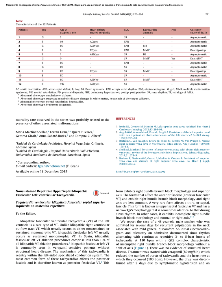
Documento descargado de http://www.elsevier.es el 19/11/2016. Copia para uso personal, se prohíbe la transmisión de este documento por cualquier medio o formato. Scientific letters / Rev Esp Cardiol. 2016;69(2):216–228 221 Table Characteristics of the 12 Patients Patients Sex Age at diagnosis, mo Heart defect/ treated surgically ECG Extracardiac anomaly PHT Outcome/ cause of death 1 G 2 - SR - - Asymptomatic 2 B 60 AC/yes EAR - - Asymptomatic 3 G PD ASD/yes EAR MR - Asymptomatic 4 B 0 TF/yes EAR MMSa - Death/postop 5 B 1 ASD/yes SR DS - Asymptomatic 6 G 0 - SR MMSb Yes Death/PHT 7 B PD - EAR - - Asymptomatic 8 G PD - SR - - Asymptomatic 9 B PD TF/yes SR MMSc - Death/postop 10 B PD - SR - - Asymptomatic 11 G PD ASD/no SR MMSd Yes Death/PHT 12 B 192 ASD/yes SR - - Asymptomatic AC, aortic coarctation; ASD, atrial septal defect; B, boy; DS, Down syndrome; EAR, ectopic atrial rhythm; ECG, electrocardiogram; G, girl; MMS, multiple malformation syndrome; MR, mental retardation; PD, prenatal diagnosis; PHT, pulmonary hypertension; postop, postoperative; SR, sinus rhythm; TF, tetralogy of Fallot. a Abnormal phenotype, omphalocele, diabetes. b Abnormal phenotype, suspected metabolic disease, changes in white matter, hypoplasia of the corpus callosum. c Abnormal phenotype, mental retardation, hypospadias. d Abnormal phenotype, brainstem dysgenesis. mortality rate observed in the series was probably related to the presence of other associated malformations. a b, b Maria Martı́nez-Villar, Ferran Gran, * Queralt Ferrer, Gemma Giralt,b Anna Sabaté-Rotés,b and Dimpna C. Albertb a Unidad de Cardiologı´a Pediátrica, Hospital Vega Baja, Orihuela, Alicante, Spain b Unidad de Cardiologı´a, Hospital Universitario Vall d’Hebron, Universidad Autónoma de Barcelona, Barcelona, Spain * Corresponding author: E-mail address: [email protected] (F. Gran). REFERENCES 1. Irwin RB, Greaves M, Schmitt M. Left superior vena cava: revisited. Eur Heart J Cardiovasc Imaging. 2012;13:284–91. 2. Angoletti G, Annecchino F, Preda L, Borghi A. Persistence of the left superior caval vein: can it potentiate obstructive lesions of the left ventricle? Cardiol Young. 1999;9:285–90. 3. Bartram U, Van Praagh S, Levine JC, Hines M, Bensky AS, Van Praagh R. Absent right superior vena cava in visceroatrial situs solitus. Am J Cardiol. 1997;80: 175–83. 4. Sheik AS, Mazhar S. Persistent left superior vena cava with absent right superior vena cava: review of the literature and clinical implications. Echocardiography. 2014;31:674–9. 5. Badessa F, Pizzimenti G, Grasso P, Merlino A, Vasquez L. Persistent left superior vena cava and absence of right superior vena cava. Ital Heart J Suppl. 2003;4:424–7. Available online 18 December 2015 http://dx.doi.org/10.1016/j.rec.2015.10.002 Nonsustained Repetitive Upper Septal Idiopathic Fascicular Left Ventricular Tachycardia form exhibits right bundle branch block morphology and superior axis. The forms that affect the anterior fascicle (anterior fascicular VT) and exhibit right bundle branch block morphology and right axis are less common. A very rare form affects a third, or septal, fascicle. This form is known as upper septal fascicular VT and has a narrow QRS morphology that is sometimes identical to that during sinus rhythm. In other cases, it exhibits incomplete right bundle branch block morphology and normal or right axis.3–5 We report the case of a 48-year-old male smoker who was admitted for several days for recurrent palpitations in the neck associated with mild general discomfort. An initial electrocardiogram and telemetry on admission documented sinus rhythm alternating with continuous repetitive 3- to 5-beat bursts of tachycardia at 110 bpm with a QRS complex characteristic of incomplete right bundle branch block morphology without a shift of axis (Figure 1). There was no evidence of structural heart disease. Treatment was started with verapamil (80 mg/8 h), which reduced the number of bursts of tachycardia and the heart rate at which they occurred (100 bpm). However, the drug was discontinued after 2 days due to symptomatic hypotension and an Taquicardia ventricular idiopática fascicular septal superior izquierda no sostenida repetitiva To the Editor, Idiopathic fascicular ventricular tachycardia (VT) of the left ventricle is a rare type of VT. Unlike idiopathic right ventricular outflow tract VT, which usually occurs as either nonsustained or sustained monomorphic VT, idiopathic fascicular left VT usually occurs as sustained monomorphic VT. In Spain, idiopathic fascicular left VT ablation procedures comprise less than 10% of all idiopathic VT ablation procedures.1 Idiopathic fascicular left VT is commonly seen in verapamil-sensitive patients without structural heart disease. The mechanism of this tachycardia is reentry within the left-sided specialized conduction system. The most common form of these tachycardias affects the posterior fascicle and is therefore known as posterior fascicular VT.2 This Documento descargado de http://www.elsevier.es el 19/11/2016. Copia para uso personal, se prohíbe la transmisión de este documento por cualquier medio o formato. 222 Scientific letters / Rev Esp Cardiol. 2016;69(2):216–228 Figure 1. A: 12-lead electrocardiogram of the patient; B: ECG telemetry. electrophysiologic study was performed without drugs. A Hisventricular interval of 53 ms in sinus rhythm was observed, with a constant His-ventricular-atrial sequence and a His-ventricular interval of 8 ms during the repetitive nonsustained tachycardia complex (Figure 2A). The CARTOW 3 system was used to guide an irrigated-tip ablation catheter into the left ventricle using an atrial transseptal approach. Ventricular activation mapping of the Purkinje potentials recorded at QRS onset was performed, with visualization of the anterior and posterior fascicles of the left bundle branch (Figure 2B). At a site adjacent to the branching point of the anterior and posterior fascicles, the interval between the fascicular potential and QRS onset was virtually identical during sinus rhythm and tachycardia (32 ms) (Figure 2C), suggesting that this part of the conduction system was involved in the tachycardia.4 Furthermore, during activation mapping of this site, the tachycardia changed from repetitive nonsustained tachycardia to sustained tachycardia with occasional interruptions; the sustained tachycardia was reinduced by ventricular pacing. At this point in the procedure, it was possible to entrain the tachycardia. A difference was obtained between the post-pacing interval and tachycardia cycle length (0 ms), with an interval between the peak pacing level and the QRS onset equal to the interval between the fascicular potential and QRS onset. Nevertheless, there was a certain degree of fusion, which may have been due to capture from the adjacent ventricle (Figure 2D). The findings suggested that there was a reentry mechanism and that the ablation catheter was positioned at the site of the tachycardia circuit. Three radiofrequency ablations were performed at this site (Figure 2B) during sinus rhythm (up to 40 W, 15 mL/h irrigation). Following this procedure, the tachycardia could not be reinduced, stable sinus rhythm without bursts of VT was maintained, and baseline QRS morphology remained unchanged (Figure 2E). After a follow-up of 3 months without drugs, the patient was asymptomatic without new episodes of tachycardia. Upper septal idiopathic fascicular VT is very rare. This tachycardia uses portions of the posterior fascicle as the anterograde limb (which can be considered as an orthodromic form of posterior fascicular VT5) and the septal fascicle as the retrograde limb, with simultaneous passive activation of the right bundle branch and anterior fascicle, which accounts for the relatively narrow QRS that can be very similar to baseline QRS. This form of tachycardia is often associated with a history of ablation of a typical posterior fascicular VT,2,3,5 although our patient had not undergone this procedure. To our knowledge, this case is unique in the literature, given the electrocardiographic presentation of repetitive nonsustained tachycardia. Documento descargado de http://www.elsevier.es el 19/11/2016. Copia para uso personal, se prohíbe la transmisión de este documento por cualquier medio o formato. Scientific letters / Rev Esp Cardiol. 2016;69(2):216–228 A 223 B 50 ms III aVL Successful ablation site V1 V3 Anterior fascicle His RA Proximal His HV 8 ms HV 53 ms Distal His aRV Posterior fascicle D C III I aVL II V1 aVF V3 FP-QRS 32 ms V1 FP-QRS 32 ms V2 dAbl V5 V6 pAbl 575 ms dAbl 575 ms RA aRV aRV S1 E S1 550 550 S1 I aVR V1 V4 II aVL V2 V5 III aVF V3 V6 II Figure 2. A: Intracavity recording and electrocardiographic leads of 1 (left) sinus beat and 1 tachycardia beat (right); B: Right anterior oblique view of the electroanatomic reconstruction and activation map of the fascicular potential created with the CARTOW 3 system; C: Intracavity recordings of the ablation catheter at the correct site during 1 sinus beat (left) and 1 tachycardia beat (right); D: Detail of tachycardia entrainment from the successful ablation site; E: 12-lead electrocardiogram after tachycardia ablation. dAbl, distal ablation; pAbl, proximal ablation; RA, right atrium; aRV, apex of the right ventricle; HV, His-ventricular; FP, fascicular potential. Documento descargado de http://www.elsevier.es el 19/11/2016. Copia para uso personal, se prohíbe la transmisión de este documento por cualquier medio o formato. 224 Scientific letters / Rev Esp Cardiol. 2016;69(2):216–228 Miguel A. Arias,* Alberto Puchol, Marta Pachón, Finn Akerström, and Luis Rodrı́guez-Padial 2. Unidad de Arritmias y Electrofisiologı́a Cardiaca, Servicio de Cardiologı´a, Hospital Virgen de la Salud, Toledo, Spain 3. * Corresponding author: E-mail address: [email protected] (M.A. Arias). 4. Available online 23 December 2015 5. REFERENCES 1. Ferrero de Loma-Osorio A, Gil-Ortega I, Pedrote-Martı́nez A; Spanish Catheter Ablation Registry collaborators. Spanish Catheter Ablation Registry. 13th Official Heart Team Decision-making: Democracy or Dictatorship? Toma de decisiones por el equipo cardiaco: democracia o dictadura? ? To the Editor, There are large groups of cardiological patients with no strong evidence base to guide medical actions. Recently, the difficulty of clinical decision-making has increased considerably due to several factors such as the increase in life expectancy and comorbidities, the lack of evidence derived from clinical trials in certain subgroups, or respect for patient preferences. Heart team decision-making1 has arisen in response to the challenge of complex cases and has received the strongest recommendations. However, this system of decision-making has not been scientifically evaluated. It is very difficult to conduct specific experiments to identify errors in clinical decision-making, and therefore the epistemological basis supporting heart team decision-making is weak. However, this barrier can be crossed with the methodological tools of social sciences that have already been used to study group http://dx.doi.org/10.1016/j.rec.2015.10.013 decision-making in other fields such as business or electoral systems. The aim of this article was to assess heart team decisionmaking from the perspective of social sciences. Study of the aggregation of individual preferences is important because in many human activities, decisions are collectively taken. Nicolas de Condorcet, an eighteenth-century French mathematician and revolutionary, pioneered the application of probability theory to collective decision-making. One of his objectives was to mathematically substantiate decision-making in democratic systems, which began to be instituted in the late eighteenth century. Condorcet’s Jury Theorem2 concluded that if each member of a jury is more likely than not to make a correct decision, the probability that the highest vote of the jury is the correct decision increases as the number of members of the jury increases. This theorem also states that if there are only 2 options, the overall preferences of a group are consistent with the preferences of the majority of its members. Therefore, heart team decision-making has emerged as an attractive tool for decision-making in complex patients. However, when there are more than 2 options, the general preference of a group cannot match the preferences of its members3; this might occur, for example, in a heart team deciding B A First preference Second preference Heart team member 1 CABG PCI Medical treatment Heart team member 2 PCI Medical treatment CABG Heart team member 3 Report of the Spanish Society of Cardiology Working Group on Electrophysiology and Arrhythmias (2013). Rev Esp Cardiol. 2014;67:925–35. Liu Y, Fang Z, Yang B, Kojodjojo P, Chen H, Ju W, et al. Catheter ablation of fascicular ventricular tachycardia: long term clinical outcomes and mechanisms of recurrence. Circ Arrhythm Electrophysiol. 2015 Sept 18. http://dx.doi.org/ 10.1161/CIRCEP.115.003080. Sung RK, Kim AM, Tseng ZH, Han F, Inada K, Tedrow UB, et al. Diagnosis and ablation of multiform fascicular tachycardia. J Cardiovasc Electrophysiol. 2013;24:297–304. Ma W, Wang X, Cingolani E, Thajudeen A, Gupta N, Nageh MF, et al. Mapping and ablation of ventricular tachycardia from the left upper fascicle. How to make the most of the fascicular potential. Circ Arrhythm Electrophysiol. 2013;6:e47–51. Talib AK, Nogami A, Nishiuchi S, Kowase S, Kurosaki K, Matsui Y, et al. Verapamilsensitive upper septal idiopathic left ventricular tachycardia: prevalence, mechanism, and electrophysiological characteristics. J Am Coll Cardiol EP. 2015. http://dx.doi.org/10.1016/j.jacep.2015.05.011. Medical treatment CABG CABG Third preference Medical treatment PCI PCI Figure 1. A: Example of preferences matrix of the heart team for patients with multivessel coronary disease. B: Condorcet’s paradox showing that collective preferences can be cyclic (intransitive), even if individual preferences are not cyclic. CABG, coronary artery bypass graft; PCI, percutaneous coronary intervention.
